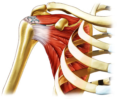
Rotator cuff repair is surgery to repair a torn tendon in the shoulder. The procedure can be done with a large (open) incision or with shoulder arthroscopy, which uses small buttonhole-sized incisions. The rotator cuff is a group of muscles and tendons that form a cuff over the shoulder joint. These muscles and tendons hold the arm in its joint and help the shoulder joint to move. The tendons can be torn from overuse or injury.
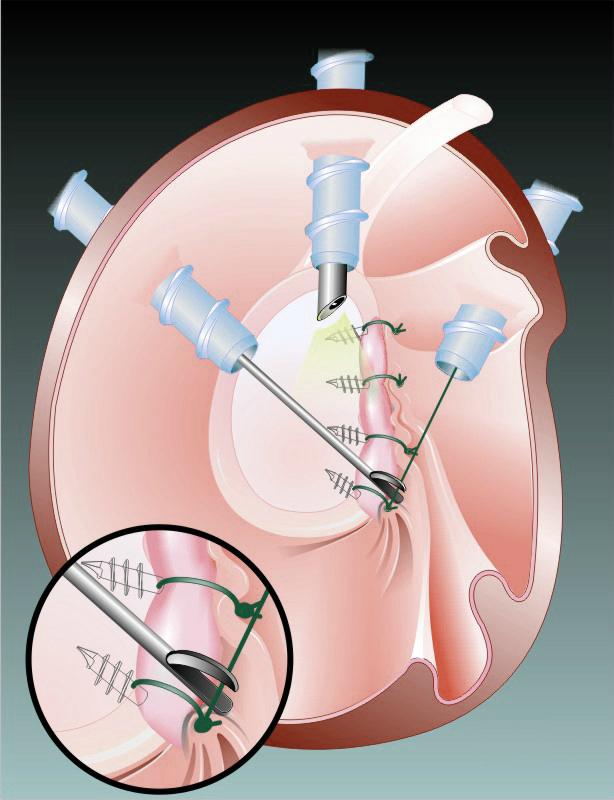
The Arthroscopic Bankart Repair is an effective procedure to treat patients that have anterior shoulder instability. The majority of patients who suffer a traumatic anterior dislocation of their shoulder will tear the fibrocartilage labrum at the front of the shoulder. Many of these patients will go on to develop recurrent instability in their shoulder and keep dislocating. This will have a significant effect on the ability to participate in sport and sometimes also their work. It is the tear in the labrum that is largely responsible for allowing their shoulder to continue to dislocate.
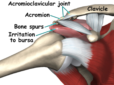
Subacromial decompression (acromioplasty) is an operation on your shoulder. It’s used to treat a condition called shoulder impingement. This is when the bones and tendons in your shoulder rub against each other when you raise your arm, causing pain. The word ‘subacromial’ means ‘under the acromion’. The acromion is part of your shoulder blade (scapula), and it helps to form your shoulder joint. If you’re having a subacromial decompression procedure it will probably be done through keyhole surgery (arthroscopy) under a general anaesthetic. Most people get to go home on the same day. Find out more about having the procedure in our section ‘what happens during subacromial decompression’ below.
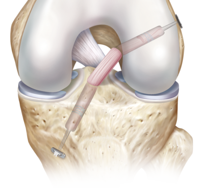
ACL Reconstruction ACL is the abbreviation for the Anterior Cruciate Ligament. This is one of the major stabilising ligaments within the knee. It connects the femur (thigh bone) to the tibia (leg bone) and prevents abnormal movement (instability) occurring between the two. More specifically it provides rotatory stability to the knee to allow movements such as pivoting or sudden change in direction to occur without the knee giving way.

PCL Reconstruction The posterior cruciate ligament, or PCL, is the strongest ligament of the knee. While the anterior cruciate ligament, or ACL is injured more often than the PCL and is more commonly discussed, PCL injuries account for more than 20% of reported knee injuries. Because the ACL sits just in front of the PCL, injuries to the PCL are commonly missed and left undiagnosed. PCL injuries are classified according to the amount of injury to the functional ligament: • Grade I: partial PCL tear • Grade II: near complete PCL tear • Grade III: a complete PCL tear – the ligament is non-functional
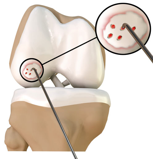
Articular or hyaline cartilage is the tissue lining the surface of two bones in a joint. It helps the bones to move smoothly against each other and can withstand the pressure of activities such as running and jumping. Articular cartilage does not have a direct blood supply so it has less capability of repairing by itself and can lead to degeneration of the articular surface (osteoarthritis). Chondroplasty refers to the surgical repair of torn or damaged cartilage. It is usually performed as day-case surgery through arthroscopy. An arthroscopic procedure involves the insertion of a telescope through a small incision, allowing your surgeon to clearly view the operating site on a monitor without having to make a large opening. Along with the arthroscope, other surgical instruments are inserted to repair the damage.